Focused Ultrasound Neuromodulation: Can Sound Waves Precisely Modulate the Human Brain?
 For decades, psychiatrists and neurologists have tried to influence dysfunctional brain circuits using medications, electrical stimulation, magnetic stimulation and implanted electrodes.
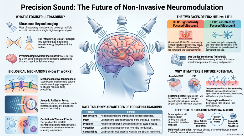
Focused ultrasound (FUS) introduces a very different possibility: using precisely directed sound waves to influence activity in selected areas of the brain—potentially even deep structures—without making an incision.
Depending on the intensity used, focused ultrasound can do two very different things:
- High-intensity focused ultrasound (HIFU) can permanently destroy a precisely selected area of tissue.
- Low-intensity focused ultrasound (LIFU) can potentially alter neuronal activity temporarily and reversibly.
The latter is particularly interesting for the future of psychiatry and precision neuromodulation.
A major review in Neurotherapeutics describes focused ultrasound as a non-invasive, steerable technology capable of concentrating energy within highly restricted regions of the nervous system, with potential applications ranging from lesioning to reversible neuromodulation.
What Is Focused Ultrasound?
Ultrasound is simply sound at frequencies above the normal range of human hearing.
Medical ultrasound systems generate these waves through piezoelectric transducers, which convert electrical energy into mechanical vibrations.
With conventional ultrasound, these waves are primarily used to create images.
With focused ultrasound, however, multiple acoustic waves can be timed so that they converge at a particular location.
The principle is similar to concentrating sunlight with a magnifying glass.
Instead of concentrating light, focused ultrasound concentrates mechanical acoustic energy.
Modern phased-array systems can alter the timing of individual transducers to steer the focal point electronically.
The result is an intriguing possibility:
deliver significant energy to a very small area deep inside the brain while exposing surrounding tissue to much less energy.
HIFU: Creating a Brain Lesion Without Open Surgery
At high intensities, focused ultrasound produces substantial heating.
When temperatures become sufficiently high, proteins denature and tissue is permanently damaged. The review describes progressive tissue injury in approximately the 43–60°C range, depending on both temperature and exposure duration.
This forms the basis of high-intensity focused ultrasound, or HIFU.
Rather than inserting a surgical probe through the skull, acoustic energy from outside the head can converge on a selected deep-brain target.
MR-Guided Focused Ultrasound
Modern systems commonly combine HIFU with MRI.
Magnetic resonance-guided focused ultrasound, or MRgFUS, allows clinicians to monitor temperature changes in real time using MR thermometry.
This helps physicians estimate the evolving lesion while limiting unwanted heating of surrounding structures.
Focused-ultrasound thalamotomy became an important clinical application in conditions such as essential tremor, demonstrating that clinically meaningful functional neurosurgery can be performed without conventional cranial surgery.
LIFU: Modulating the Brain Without Destroying Tissue
Low-intensity focused ultrasound is fundamentally different.
Instead of creating a permanent lesion, LIFU attempts to change neuronal activity reversibly.
Experimental studies have reported both:
- increased neuronal firing, and
- suppression of neuronal activity.
Regions studied have included the motor cortex, sensory cortex, visual cortex, hippocampus, thalamus and brainstem. Changes have also been demonstrated using EEG and evoked potentials.
This combination of properties makes LIFU particularly interesting:
non-invasive + deep-reaching + focal + potentially reversible.
That is a combination few existing neuromodulation technologies can currently provide.
How Does Ultrasound Change Neuronal Activity?
This remains one of the major scientific uncertainties.
Ultrasound is a mechanical wave, and several mechanisms have been proposed.
Mechanosensitive Ion Channels
Neuronal membranes contain proteins that respond to mechanical forces.
Ultrasound may deform cellular membranes sufficiently to influence these mechanosensitive channels.
Experimental work reviewed in the paper describes effects involving voltage-gated calcium, sodium and potassium channels following ultrasound exposure.
Alterations in these channels could change the probability that a neuron fires.
Acoustic Radiation Force
Sound waves transfer momentum as they move through tissue.
That mechanical pressure may exert microscopic forces upon membranes and cellular structures, potentially influencing neuronal excitability.
Cavitation
Ultrasound can also affect microscopic gas bubbles.
At certain intensities, these bubbles may oscillate or collapse.
More violent cavitation is especially relevant to high-intensity ultrasound because it can contribute to tissue destruction.
Thermal Effects
Even low-intensity ultrasound may create very small temperature changes.
These are far below the levels used to create a lesion, but neuronal membranes and ion channels are themselves sensitive to temperature.
Consequently, the distinction between “mechanical” and “thermal” neuromodulation may not always be absolute.
The review ultimately concludes that several mechanisms may contribute simultaneously, and their relative importance remains uncertain.
A Fascinating Problem: Is Ultrasound Really Stimulating the Intended Brain Region?
Early ultrasound neuromodulation experiments produced another scientific surprise.
Some apparently direct brain responses may actually have resulted from activation of the auditory system.
Ultrasound transmitted through the skull can stimulate the cochlea and auditory pathways, producing widespread neuronal responses that may be mistaken for focal brain stimulation.
This is extremely important experimentally.
If EEG activity changes after ultrasound stimulation, researchers must demonstrate that the effect actually arose from the intended brain target rather than an indirect sensory pathway.
However, auditory activation does not adequately explain all reported effects, particularly some observations of suppression of evoked potentials and neuronal activity.
The mechanism of LIFU therefore remains an active area of research.
Focused Ultrasound vs TMS
Transcranial magnetic stimulation has already established that psychiatric disorders can be treated by modifying neural circuits from outside the skull.
But TMS has a physical limitation.
Its strongest effects are relatively close to the cortical surface.
Focused ultrasound offers the theoretical possibility of reaching much deeper structures while retaining high anatomical precision.
The review compares the approximate spatial resolution of conventional TMS with focused ultrasound and notes that FUS can potentially achieve millimetre or even submillimetre-scale focusing.
That difference could become clinically significant.
Many important psychiatric circuits involve structures deeper than the surface cortex.
Examples include networks involving the:
- anterior cingulate cortex,
- insula,
- striatum,
- thalamus,
- amygdala,
- hippocampus, and
- deeper components of cortico-striato-thalamo-cortical circuits.
Conventional non-invasive stimulation cannot always directly reach these structures with high precision.
Focused ultrasound potentially could.
Why This Matters for Psychiatry
Psychiatric disorders are increasingly being understood as disturbances of brain networks, rather than abnormalities of one isolated brain region.
Consider depression.
Rather than asking:
“Which part of the brain causes depression?”
modern neuroscience increasingly asks:
“Which networks regulating mood, reward, cognition and emotional processing are functioning abnormally?”
The same logic applies to conditions such as:
- obsessive-compulsive disorder,
- addiction,
- depression,
- chronic pain,
- anxiety disorders,
- and potentially other neuropsychiatric conditions.
The review specifically identifies psychiatric disorders as potential future targets for more subtle or distributed network neuromodulation.
However, an important distinction must be maintained.
LIFU is not currently an established routine treatment for psychiatric disorders.
The review itself describes many proposed applications beyond established neurological uses as promising but still speculative.
The technology is exciting precisely because of what it may eventually enable, not because every theoretical application has already been clinically proven.
Could We Stimulate Several Brain Regions at Once?
Another advantage of phased-array focused ultrasound is the possibility of multifocal stimulation.
Rather than targeting one location, future systems could potentially stimulate several nodes within a brain network.
The reviewed technology allows acoustic foci to be electronically steered and raises the possibility of creating multiple focal targets.
This could be especially important in psychiatry.
Mood, attention, motivation, impulse control and emotional regulation are not produced by isolated structures.
They emerge from communication between interconnected regions.
Future neuromodulation may therefore move away from:
one disorder → one brain region → one stimulation protocol
toward:
individual network abnormality → personalised targets → adaptive stimulation.
EEG, Brain Mapping and Closed-Loop Neuromodulation
One of the most exciting possibilities described in the review is combining focused ultrasound with physiological measurements such as:
- EEG,
- fMRI, and
- intracranial electrophysiology.
This creates the possibility of closed-loop neuromodulation.
Instead of giving every patient the same stimulation regardless of what the brain is doing, a future system could potentially:
1. Measure brain activity
↓
2. Identify an abnormal network state
↓
3. Deliver targeted focused ultrasound
↓
4. Measure the electrophysiological response
↓
5. Adjust the stimulation
This is conceptually very different from conventional fixed-dose stimulation.
Neuromodulation could eventually become responsive rather than simply repetitive.
Focused Ultrasound Can Also Open the Blood–Brain Barrier
Perhaps one of the most remarkable applications of focused ultrasound does not involve direct neuronal stimulation at all.
The brain is protected by the blood–brain barrier, which tightly regulates which molecules can enter neural tissue from the circulation.
This protects the brain—but also makes many neurological treatments difficult.
Focused ultrasound combined with microbubbles can produce temporary, localised opening of the blood–brain barrier.
In principle, this could allow future treatments to deliver:
- medications,
- antibodies,
- nanoparticles,
- biological molecules,
- or gene therapies
to selected areas of the brain.
The review therefore suggests that focused-ultrasound-assisted drug or gene delivery may ultimately prove just as important as direct neuromodulation.
Why Focused Ultrasound Is So Interesting
Few neuromodulation technologies offer this combination of properties.
Focused ultrasound can potentially be:
Non-invasive
No electrode needs to be implanted in the brain.
Highly focal
Energy can be concentrated within a relatively small anatomical region.
Deep-reaching
Targets need not be restricted to superficial cortex.
Steerable
Phased arrays allow electronic control of the focal point.
Multifocal
Multiple components of a neural network may eventually be targeted.
MRI and EEG compatible
Brain activity can potentially be measured while stimulation is delivered.
Closed-loop capable
Physiological feedback could modify stimulation dynamically.
Reversible or permanent
LIFU may temporarily alter activity, whereas HIFU can create permanent lesions.
These characteristics account for much of the scientific excitement surrounding the technology.
What We Still Do Not Know
Despite its promise, focused ultrasound—especially LIFU—still faces major unanswered questions.
We need better evidence regarding:
- the precise cellular mechanism of neuromodulation,
- parameters that produce excitation versus inhibition,
- individual differences caused by skull anatomy,
- optimal stimulation frequency and intensity,
- duration of treatment effects,
- long-term safety of repeated stimulation,
- clinically meaningful targets,
- and whether physiological biomarkers can reliably guide treatment.
The review emphasises that low-intensity ultrasound remains less developed than high-intensity lesioning and requires both technological refinement and greater mechanistic clarity.
This distinction matters particularly in psychiatry.
A promising neuroscience technology is not automatically an established psychiatric treatment.
Evidence must come before clinical enthusiasm.
From Treating Diagnoses to Modulating Circuits
Focused ultrasound ultimately represents a broader transformation occurring in psychiatry and neuroscience.
Traditional psychiatry largely classified illness according to symptoms.
Modern neuroscience increasingly asks whether those symptoms can also be understood through abnormalities in:
- neural circuits,
- functional connectivity,
- electrophysiological activity,
- cognition,
- and network regulation.
Neuromodulation technologies such as TMS, DBS and focused ultrasound, together with tools including EEG, quantitative EEG, neuroimaging and cognitive testing, are helping move the field toward this more objective and circuit-based understanding of brain disorders.
Focused ultrasound may ultimately become particularly valuable because it could provide something that has historically been difficult to achieve:
precise, non-invasive access to deep neural circuits.
The original review concludes that focused ultrasound has the potential to become a precise, flexible, non-invasive and clinically translatable neuromodulation platform, while emphasising that technological refinement and stronger understanding of its mechanisms remain necessary.
For psychiatry, the potential is substantial.
TMS demonstrated that psychiatric symptoms can improve when selected neural circuits are modified.
Focused ultrasound raises the next question:
What becomes possible when we can reach those circuits deeper, more precisely and perhaps eventually in a closed-loop, personalised manner?
About Dr. Srinivas Rajkumar T
Dr. Srinivas Rajkumar T, MBBS, MD (AIIMS New Delhi), DNB, MBA, is a Senior Consultant Psychiatrist with Apollo Hospitals and Assistant Professor of Psychiatry at Sri Balaji Medical College & Hospital, Chennai.
His clinical and academic interests include biological psychiatry, interventional psychiatry, ADHD and attention disorders, quantitative EEG, cognitive assessment, neurofeedback and objective approaches to understanding brain function.
His approach combines detailed psychiatric evaluation with contemporary methods of assessing cognition and brain function where clinically appropriate.
Consultation in Chennai
Dr. Srinivas Rajkumar T
Senior Consultant Psychiatrist
Apollo Clinic
Opp. Phoenix Market City
Velachery, Chennai
Appointments: +91 85951 55808
Email: srinivasaiims@gmail.com
Focused ultrasound neuromodulation for psychiatric disorders remains largely investigational and is discussed here as an emerging neuroscience technology rather than as a currently offered clinical treatment.
Reference
Darrow DP. Focused Ultrasound for Neuromodulation. Neurotherapeutics. 2019;16:88–99. Published online 28 November 2018