EEG Microstates and Large-Scale Brain Networks: Understanding the Brain’s Hidden Dynamics
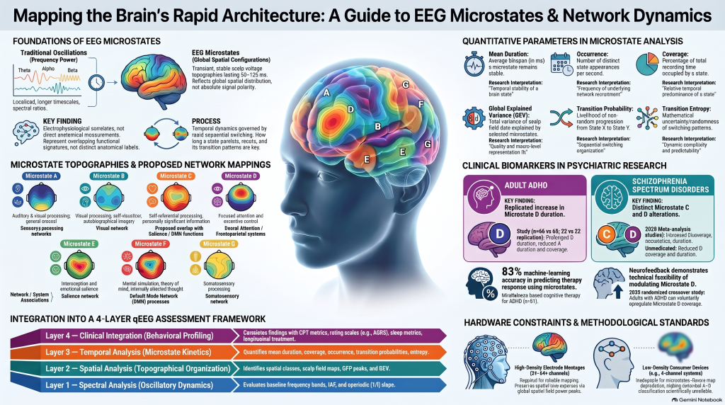 How quantitative EEG is helping neuroscience move beyond brain waves towards understanding the rapidly changing networks underlying attention, thinking, emotions and psychiatric disorders.
Introduction: The Brain Is Never Truly at Rest
Even when we sit quietly with our eyes closed, the brain is extraordinarily active.
Thoughts emerge and disappear. Memories surface unexpectedly. Attention shifts between the outside world and our internal experiences. The mind may wander into the past, anticipate the future or briefly settle into focused awareness.
These experiences involve complex interactions between billions of neurons distributed across different brain regions.
For decades, electroencephalography (EEG) has helped us examine this activity through electrical oscillations commonly known as delta, theta, alpha, beta and gamma waves.
However, the brain is much more than a collection of oscillations.
Different brain regions continuously coordinate their activity, forming large-scale functional networks. These networks do not remain static. They reorganize dynamically, sometimes within fractions of a second.
A particularly fascinating approach to studying these rapid changes is known as EEG microstate analysis.
This field offers a new perspective on how brain activity is organized in time and space—and potentially how that organization changes in psychiatric and neurological disorders.
1. Moving Beyond Traditional qEEG
Quantitative electroencephalography (qEEG) involves applying mathematical and computational techniques to recorded EEG signals.
Traditional qEEG analysis often examines:
- Spectral power: How much electrical activity is present in different frequency bands?
- Alpha peak frequency: What is the dominant frequency of posterior alpha activity?
- Asymmetry: Are there differences in electrical activity between corresponding regions of the brain?
- Connectivity: How are electrical signals across different regions statistically related?
- Event-related activity: How does the brain respond to specific stimuli or cognitive tasks?
These measurements provide valuable information about brain physiology.
However, conventional frequency-based analysis does not fully describe how the spatial organization of electrical activity changes from one moment to the next.
Consider two individuals with similar resting alpha power.
Their spectral profiles may appear comparable, yet their brains could differ in how frequently particular electrical configurations occur, how long those configurations persist and how rapidly they transition between them.
Microstate analysis attempts to characterize this additional dimension.
Traditional qEEG helps describe the frequencies of brain activity. Microstate analysis examines the rapidly changing spatial configurations of that activity.
The approaches are complementary rather than competing.
2. What Are EEG Microstates?
An EEG microstate is a brief period during which the spatial pattern of electrical potentials across the scalp remains approximately stable, even while signal amplitudes continue to fluctuate.
These periods commonly last around 50–120 milliseconds.
In other words, during a typical second of EEG recording, the brain may pass through several distinct configurations of electrical activity.
Interestingly, researchers have observed that certain spatial patterns recur repeatedly across individuals.
These recurring patterns are called microstate classes.
The concept is sometimes described metaphorically as the brain’s atoms of thought.
This is an evocative description, but it should not be interpreted literally. A microstate does not represent an identifiable thought, emotion or memory. Instead, it reflects a short-lived configuration of large-scale electrical activity.
The four classical microstates
Early research identified four frequently recurring configurations, conventionally labeled:
Microstate A, Microstate B, Microstate C and Microstate D.
These are distinguished primarily by the distribution of positive and negative electrical potentials across the scalp.
Subsequent research has explored additional configurations, including microstates E, F and G.
The functional significance of individual microstates remains an active research question, but evidence suggests relationships with different aspects of information processing.
For example:
| Microstate | Proposed functional association |
|---|---|
| A | Auditory and visual processing, arousal |
| B | Visual imagery and autobiographical processing |
| C | Self-referential processing and personally significant information |
| D | Attention and executive functioning |
| E | Emotional and interoceptive salience |
| F | Default mode network-related processing |
| G | Somatosensory processing |
These associations are derived from studies combining EEG, cognitive tasks, brain source analysis and functional neuroimaging.
Importantly, they are not fixed anatomical assignments. Different studies have produced partially different mappings, and the functional meaning of a microstate depends on the methods used to identify it.
A systematic review by Tarailis and colleagues, published in Brain Topography in 2024, provides a particularly useful synthesis of this literature.
3. Understanding the Brain’s Large-Scale Networks
The human brain does not function as a collection of independent anatomical compartments.
Thinking, attention, emotion and behavior emerge from interactions among distributed brain regions.
Functional neuroimaging has identified several large-scale networks that participate in these processes.
Three networks are especially relevant to psychiatry.
The Default Mode Network (DMN): The Internally Directed Brain
The Default Mode Network includes regions such as the medial prefrontal cortex, posterior cingulate cortex, precuneus and angular gyrus.
It is frequently associated with:
- Self-referential thinking
- Autobiographical memory
- Mental simulation of future events
- Internally directed attention
- Spontaneous thought and mind-wandering
The DMN does not simply represent an inactive or distracted brain.
It performs important cognitive functions, particularly those related to our internal mental world.
Problems may arise when the coordination between internally oriented processing and external task demands becomes inefficient.
This has attracted particular interest in ADHD, depression and other psychiatric conditions.
The Salience Network (SN): Identifying What Matters
The Salience Network prominently involves the anterior insula and dorsal anterior cingulate cortex.
It helps identify information that is important to the individual, whether that information arises from the external environment or the body.
For example, a sudden loud noise, an emotionally significant facial expression or an unexpected bodily sensation may demand attention.
The salience network participates in coordinating responses to such events and interactions with other networks.
Disturbances involving salience processing have been investigated in schizophrenia, anxiety disorders, depression and several other psychiatric conditions.
The Frontoparietal Control Network (FPN): Executive Regulation
The Frontoparietal Control Network involves regions of the prefrontal and parietal cortices.
It supports functions such as:
- Working memory
- Goal-directed behavior
- Cognitive flexibility
- Problem-solving
- Executive control
Related dorsal attention systems support the selection and maintenance of attention toward task-relevant information.
These systems interact with the default mode and salience networks, depending on the individual’s cognitive demands.
For effective functioning, the brain must coordinate these systems flexibly.
The efficiency of cognition depends not merely on the activity of individual networks, but also on how their activity is coordinated over time.
4. How Are EEG Microstates Related to These Networks?
This is one of the most interesting questions in contemporary EEG research.
Functional MRI can identify distributed networks through correlations in blood-oxygen-level-dependent signals. However, this measurement reflects relatively slow physiological changes.
EEG, in contrast, measures electrical activity with millisecond temporal resolution.
Could rapidly changing EEG microstates reflect some of the electrical processes associated with the networks identified by functional MRI?
An influential study by Britz and colleagues, published in NeuroImage in 2010, investigated this question using simultaneous EEG and functional MRI.
The researchers identified relationships between classical EEG microstates and distributed resting-state networks involved in auditory processing, visual imagery, attention and interoceptive processing.
The findings provided important evidence linking rapid EEG topographical dynamics with large-scale functional organization.
Interestingly, the original study did not identify a clear default mode network correlate among the four classical microstates.
Subsequent studies using alternative clustering strategies, additional microstate classes and source-localization methods have explored possible DMN-related signatures.
This is an important scientific distinction.
An EEG microstate is not the same thing as a functional MRI network.
Microstates are spatial patterns measured at the scalp. Large-scale functional networks are distributed systems inferred through different neuroimaging and analytical techniques.
Their relationships are meaningful, but not necessarily one-to-one.
Consequently, a clinician cannot simply identify microstate D and conclude that the dorsal attention network is functioning normally or abnormally.
The interpretation requires considerably more care.
5. What Does Microstate Analysis Actually Measure?
Microstate analysis does more than identify recurring scalp patterns.
It also examines their temporal characteristics.
Four important parameters are particularly useful.
A. Mean Duration
How long does a particular microstate persist before the configuration changes?
A longer duration means that the same spatial configuration remains dominant for a longer period.
This may reflect differences in temporal stability, but its functional significance depends on the microstate, population and recording conditions.
B. Occurrence
How frequently does a particular microstate appear?
Occurrence is commonly expressed as the number of appearances per second.
C. Coverage
What percentage of the recording is occupied by a particular microstate?
For example, a microstate occupying 30% of the analyzed recording has greater temporal coverage than one occupying 15%.
Coverage does not directly measure the intensity of a particular cognitive function.
D. Transition Probability
How frequently does the brain move from one microstate class to another?
Does microstate A tend to transition into B, C or D?
Are some transitions more frequent than expected?
Such analyses examine what researchers call microstate syntax, or the sequential organization of microstates.
More advanced approaches assess transition entropy, sequence complexity and non-random temporal organization.
Together, these parameters offer a way to describe the rapidly changing architecture of scalp electrical activity.
6. EEG Microstates in ADHD: A New Understanding of Attention?
Attention-deficit/hyperactivity disorder is increasingly understood as involving difficulties in the regulation of attention, motivation, inhibitory control, working memory and other executive functions.
No single brain region explains the entire disorder.
Instead, research has implicated interactions among frontoparietal control systems, attention networks, salience processing systems, subcortical circuits and the default mode network.
A longstanding hypothesis proposes that some attentional difficulties may involve inefficient regulation of internally directed processing during tasks requiring sustained external attention.
Microstate analysis offers one possible approach to studying the temporal organization underlying these difficulties.
What does the research show?
A study by Férat and colleagues, published in Biological Psychiatry: Cognitive Neuroscience and Neuroimaging in 2022, examined resting-state microstates in adults with ADHD.
The investigators compared 66 adults with ADHD against 66 controls and used another independent dataset of 22 patients and 22 controls for validation.
A particularly interesting finding was that microstate D exhibited longer mean duration in the ADHD groups.
The spatial configurations themselves were broadly comparable between patients and controls.
This suggests that differences in ADHD may involve the temporal behavior of some recurring electrical configurations rather than an entirely different collection of configurations.
A further systematic review published by Berchio and colleagues in 2025 examined 13 studies involving ADHD and microstate analysis.
The review identified preliminary evidence for changes in microstates C and D, along with task-related abnormalities involving attention and response inhibition.
These observations are scientifically interesting, but they should not be overinterpreted.
A prolonged microstate D does not establish ADHD, identify a particular ADHD subtype or directly measure executive dysfunction in an individual patient.
Moreover, resting-state microstate patterns can be influenced by vigilance, sleep disturbance, medication effects and methodological choices.
Why does this matter clinically?
ADHD is not simply a condition of having insufficient attention.
Many individuals with ADHD can sustain intense concentration in certain situations while struggling to regulate attention in others.
A more sophisticated understanding of ADHD may therefore involve examining the flexibility and stability of underlying brain processes.
Microstate research may eventually help investigate questions such as:
- Are particular brain configurations excessively persistent during rest?
- Are transitions between electrical configurations altered during sustained-attention tasks?
- Do stimulant medications change microstate dynamics?
- Can changes in microstate parameters predict cognitive improvement?
- Are particular electrophysiological patterns associated with different symptom profiles?
These remain research questions rather than established clinical applications.
7. Microstates in Schizophrenia, Depression and Dementia
The relevance of EEG microstates extends beyond ADHD.
Schizophrenia and Psychosis
Schizophrenia has been associated with disturbances in the integration and coordination of distributed brain activity.
Microstate research has repeatedly identified abnormalities involving particular canonical configurations.
A 2025 systematic review and meta-analysis examining 24 studies found increased coverage and occurrence of microstate C across several groups along the psychosis spectrum.
In schizophrenia, increased duration of microstate C was also observed.
Some studies have reported reduced microstate D measures, particularly in unmedicated patients.
These observations may provide clues about altered functional organization in psychotic disorders.
However, they do not establish microstate C as a direct measure of delusions, hallucinations or pathological salience attribution.
Depression and Anxiety
Depression involves disturbances in emotional regulation, motivation, cognitive processing and self-referential thought.
Researchers have therefore investigated whether altered microstate dynamics relate to abnormal rumination, emotional processing and network regulation.
Findings remain heterogeneous.
Differences in patient populations, medication exposure, comorbid conditions and analytical protocols complicate direct comparisons.
Microstates may eventually contribute to understanding differences between clinical subgroups or responses to treatment, but current findings are not sufficiently robust for routine diagnostic use.
Dementia and Cognitive Decline
In neurodegenerative disorders, large-scale neuronal networks may become progressively disrupted.
EEG already provides useful research information about spectral slowing and changes in functional connectivity.
Microstate analysis adds the possibility of studying altered temporal organization.
Researchers are examining whether microstate changes correspond to cognitive impairment, disease severity or progression.
Future longitudinal studies may determine whether combining microstate features with traditional EEG measures, cognitive testing and other biomarkers improves clinical prediction.
At present, however, microstates cannot independently diagnose Alzheimer’s disease or distinguish reliably between dementia subtypes in routine practice.
8. Can EEG Microstates Become Psychiatric Biomarkers?
The word biomarker is frequently used in contemporary neuroscience.
However, a measurable difference between a patient group and healthy controls does not automatically become a clinically useful diagnostic biomarker.
For microstate analysis to become useful in routine psychiatric care, several requirements must be met.
Researchers must establish standardized recording and preprocessing protocols, demonstrate reproducibility across centers, account for medication and vigilance effects, and validate findings in independent clinical populations.
Most importantly, a microstate measure must show that it improves clinical decision-making beyond information already available from psychiatric assessment, symptom scales, cognitive testing and other established methods.
A biomarker that differs statistically between groups may still have substantial overlap between individual patients and controls.
Group-level neurophysiological findings should not be mistaken for diagnostic certainty in an individual patient.
This distinction is particularly important in ADHD, where diagnosis remains fundamentally clinical and requires developmental history, symptom assessment, functional impairment and appropriate differential diagnosis.
9. The Role of Artificial Intelligence and Advanced qEEG
Artificial intelligence provides an opportunity to analyze the complex information contained in EEG recordings.
Rather than examining only one parameter, computational models can combine multiple physiological and behavioral measurements.
For instance, an exploratory research model could integrate:
- EEG spectral power and individual alpha frequency
- Microstate duration, occurrence and coverage
- Microstate transition patterns
- Functional connectivity measures
- Continuous Performance Test (CPT) findings
- Clinical rating scales and treatment outcomes
The objective would not necessarily be to replace the clinician or produce a diagnosis from brain electrical activity alone.
A more clinically meaningful research goal might be to predict which patients are likely to improve with a particular intervention, or to identify objective changes associated with treatment response.
For ADHD, this could include prospective studies examining baseline measures and serial changes following medication or behavioral interventions.
However, sophisticated machine-learning models cannot compensate for poor-quality recordings, small unrepresentative datasets or insufficient external validation.
The promise lies in combining better physiological measurements with rigorous clinical science.
10. Can Consumer EEG Devices Measure Microstates?
Wearable EEG devices have made brain-signal acquisition more affordable and accessible.
This is potentially valuable for longitudinal studies and repeated measurements.
However, there is an important technical limitation.
Classical EEG microstate analysis depends on identifying spatial patterns distributed across the scalp.
A device recording from only a few frontal or temporal electrodes cannot reconstruct those whole-scalp patterns with the same reliability as a properly distributed multichannel EEG system.
Research-grade microstate studies commonly employ systems with 32, 64 or more electrodes.
Wearable devices with fewer channels may still be valuable for spectral analysis, monitoring physiological state and exploring other carefully validated EEG features.
But it would be scientifically inappropriate to assume that a four-channel headset can reliably reproduce conventional A–D microstate maps or their proposed large-scale network associations.
The number, distribution and quality of electrodes matter.
11. The Future: From Static Brain Maps to Dynamic Psychiatry
Much of twentieth-century neuropsychiatry attempted to understand psychiatric disorders by identifying abnormal brain regions, neurotransmitters or isolated physiological measurements.
Contemporary neuroscience increasingly emphasizes interactions among distributed neuronal systems.
This raises an important possibility:
Psychiatric symptoms may sometimes reflect not simply excessive or insufficient activity in a particular region, but altered coordination among multiple systems operating across different timescales.
EEG microstates provide one approach to examining this temporal organization.
Combined with spectral qEEG, neuropsychological assessment, cognitive testing, neuroimaging and appropriate computational methods, they may contribute to a more comprehensive understanding of brain function.
The goal should not be to manufacture a new brain-based label for every psychiatric diagnosis.
Instead, it should be to develop physiological measurements that help answer clinically meaningful questions.
Why do two patients with similar symptoms respond differently to treatment?
Can we identify meaningful neurophysiological changes before symptom improvement becomes obvious?
Can longitudinal brain recordings help us understand recovery, relapse or cognitive change?
And can these insights eventually make psychiatric treatment more personalized?
These are ambitious questions, and the available evidence does not yet provide definitive answers.
Nevertheless, they represent a scientifically worthwhile direction.
Conclusion: Understanding Not Just Brain Activity, but Brain Dynamics
The brain is not a static electrical organ.
It is a continually evolving system in which distributed neuronal populations interact across different spatial and temporal scales.
Traditional qEEG offers insights into oscillatory activity and its distribution.
EEG microstates add information about recurring electrical configurations, their duration and their temporal succession.
Research has demonstrated intriguing associations between these configurations, large-scale functional networks and psychiatric conditions including ADHD and schizophrenia.
However, their translation into individual diagnostic or treatment decisions remains an ongoing scientific challenge.
The real promise of microstate research is not that it can read thoughts or provide an immediate psychiatric diagnosis.
It is that it may help us understand how the brain organizes its activity over time—and whether disturbances in that organization contribute to psychopathology.
The next frontier in psychiatry may involve understanding not only where the brain is active or at what frequency, but also how its distributed activity evolves from one moment to the next.
About the Author | Advanced ADHD and qEEG Assessment in Chennai
Dr. Srinivas Rajkumar T, MD (Psychiatry, AIIMS New Delhi), DNB Psychiatry, MBA (BITS Pilani) is a Senior Consultant Psychiatrist practicing in Chennai.
His clinical and academic interests include adult ADHD, neurophysiology, dementia, cognitive disorders, neuromodulation and the integration of objective assessment technologies into evidence-based psychiatric practice.
At the Attention Clinic – Comprehensive 360° ADHD Care, the emphasis is on going beyond symptom checklists through detailed specialist-led assessment.
The clinical approach may integrate structured psychiatric evaluation, standardized ADHD rating scales, computerized attention testing including CPT, and qEEG-assisted assessment where appropriate and scientifically justified.
Treatment planning combines medication, psychological interventions, executive-function strategies and longitudinal monitoring according to individual clinical needs.
Dr. Srinivas has a particular interest in exploring how low-cost EEG technologies, artificial intelligence and repeated neurophysiological measurements can contribute to more accessible and clinically meaningful psychiatric research.
EEG microstate analysis remains primarily a research methodology and is not presented as an established stand-alone diagnostic test for ADHD or other psychiatric disorders.
Attention Clinic — Attention. Understood.
Specialist-led ADHD Assessment and Treatment in Chennai
Dr. Srinivas Rajkumar T
Senior Consultant Psychiatrist
MD Psychiatry (AIIMS, New Delhi) | DNB Psychiatry
Location: Apollo Clinic, Velachery, opposite Phoenix Marketcity, Chennai
Appointments: +91 85951 55808
Website: www.srinivasaiims.com
Learn more: Comprehensive 360° ADHD Care in Chennai